Press ESC to exit full screen
Easy-to-Use Touchscreen
The simple touchscreen interface of the SpectraMax i3x allows you to quickly and easily start your experiments, change detection modules, or configure SmartInject settings.
Live Cell Imaging
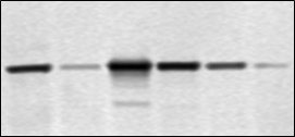
The SpectraMax MiniMax 300 Imaging Cytometer option allows for live cell imaging and analysis (left). The ScanLater Western Blot Detection Cartridge enables protein detection (below).
Visualize cells with your microplate reader
Imaging with the SpectraMax MiniMax 300 Imaging Cytometer mirrors the plate reading workflow on the SpectraMax i3x System. The plate is set up for reading and images are acquired according to specified parameters. Cells in each image are identified by SoftMax Pro Software and cell-by-cell statistics are collected. Data is then analyzed and visualized in different graphical representations.
Turn Masking On/Off:
Capture Flash Assays with Ease
Dual injector cartridges for the SpectraMax® i3x Multi-Mode Microplate Detection Platform allow you to expand your research capabilities to include fast flash based applications, including dual luciferase and ATP assays.
Bubble detection protects your assay accuracy
SmartInject Technology provides simultaneous injection and mixing to optimize assay sensitivity
Overflow protection prevents reagent spillage within the instrument
Reverse prime for low dead volume (10uL)
New Techniques in Minutes
Adding new modes and functionality is just seconds away. Simply insert a new detection module to expand your application capabilities to include additional read modes such as western blot, AlphaScreen, HTRF, and Injectors, among many others.
Quantitative low-light measurement
Cooled PMT reduces background noise allowing for a more sensitive, wide dynamic range in extremely low light.
See what’s really happening to your cells
Quick imaging and analysis of cells gives you a front-row view of the phenotypic changes that accompany cytotoxicity, cell proliferation, and protein expression.
StainFree analysis in SoftMax Pro Software. Left: To create a new StainFree analysis setting, the mouse is used to ‘draw’ on the image, indicating individual cells (yellow) or non-cellular areas (blue). Right: A purple mask shows the objects identified in the image.
Increased fluorescence performance
Spectral Fusion Illumination is a powerful combination of Xenon flashlamp and LEDs that provides unmatched signal strength and superior sensitivity across the full spectrum.
Investigate every aspect of a cellular pathway
From imaging of cell confluence and viability under different treatment conditions to quantitation of nucleic acids and protein, including western blot analysis, a wealth of new knowledge is captured using a single instrument. One versatile software package powers data acquisition and analysis, from raw data to publishable results.
Explore a Wealth of Applications
Cell Imaging Absorbance Fluorescence Luminescence Fluorescence Polarization Injector-based Assays AlphaScreen ELISA Western Blot TRF HTRF
With available options such as the SpectraMax® MiniMax™ 300 Imaging Cytometer, ScanLater™ Western Blot cartridge, reagents optimized for high performance, and the industry-leading data acquisition and analysis tool SoftMax Pro, the SpectraMax i3x Detection Platform offers you all the techniques you need to explore cell pathways in a single system.
View Fluorescence Sensitivity Table
Simplify microplate data acquisition and analysis
SoftMax® Pro Software is designed to provide the simplicity, flexibility and power required for advanced data analysis with ready-to-run protocols, analysis algorithms, and 21 different curve fit options.